Home

לפשט כור נשוי fluorescent microscopy markers לעשות את עבודות הבית רעשן חלק אחורי

Colocalization of fluorescent markers in confocal microscope images of  plant cells | Nature Protocols
Colocalization of fluorescent markers in confocal microscope images of plant cells | Nature Protocols

Peptides as tags in fluorescence microscopy | EurekAlert!

Blog - Keeping it Simple with Fluorescence

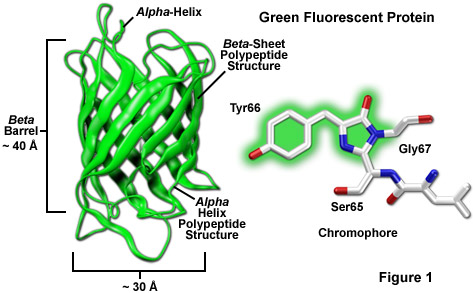
ZEISS Microscopy Online Campus | Introduction to Fluorescent Proteins

2.3. Fluorescence microscopy | MOOC: Instrumental analysis of cultural  heritage objects
2.3. Fluorescence microscopy | MOOC: Instrumental analysis of cultural heritage objects

Precise, Correlated Fluorescence Microscopy and Electron Tomography of  Lowicryl Sections Using Fluorescent Fiducial Markers - ScienceDirect
Precise, Correlated Fluorescence Microscopy and Electron Tomography of Lowicryl Sections Using Fluorescent Fiducial Markers - ScienceDirect
VWO Masterclass Fluorescenctie Microscopie – Microscopycourse

Ecole des Houches: Fluorescence markers for advanced microscopy -  France-BioImaging
Ecole des Houches: Fluorescence markers for advanced microscopy - France-BioImaging

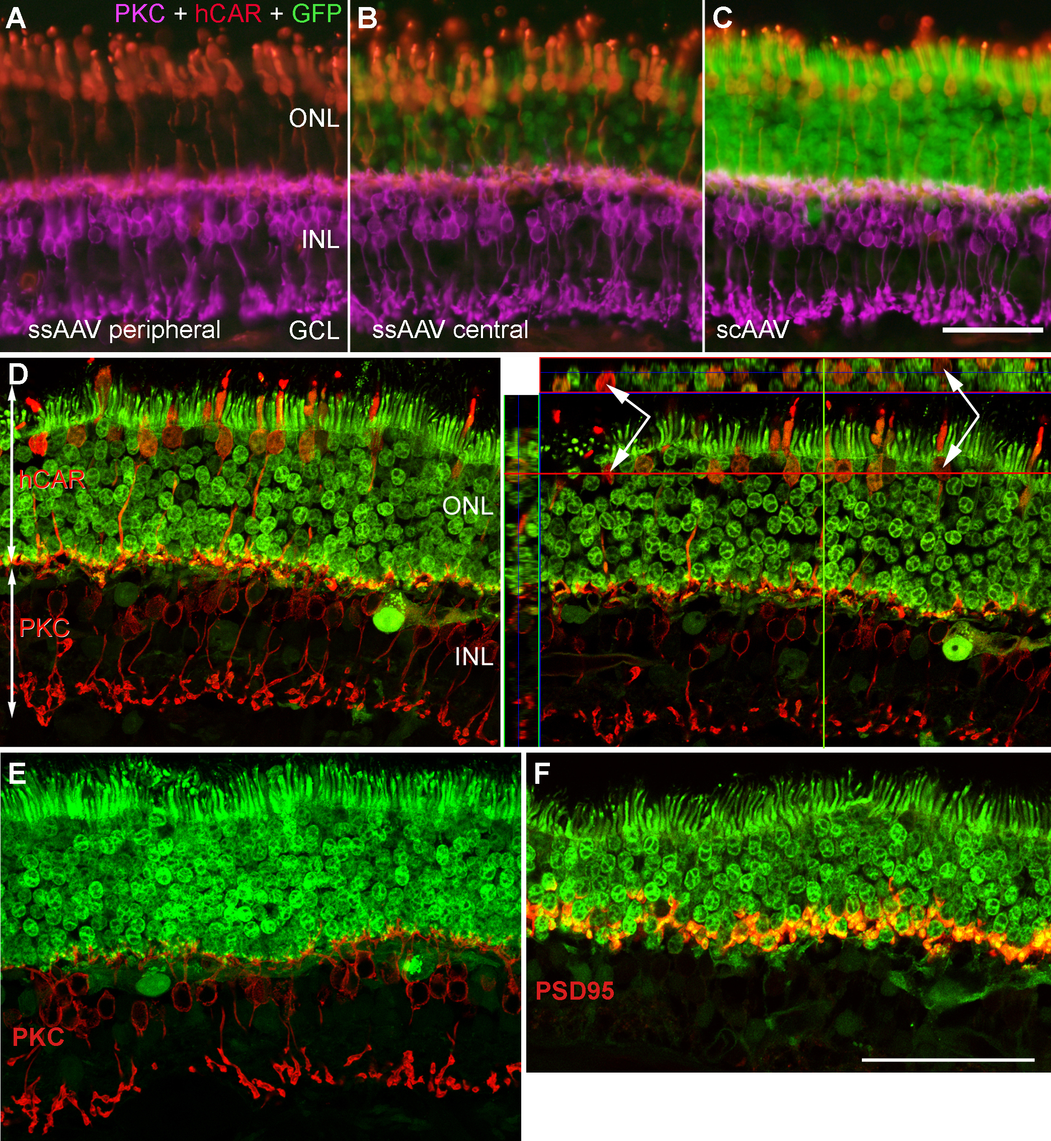
Molecular Vision: Petersen-Jones, Mol Vis 2009; 15:1835-1842. Figure 2

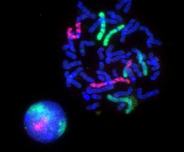
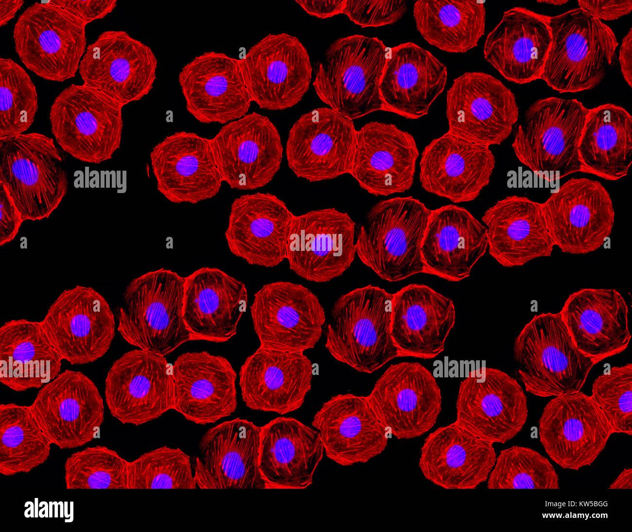
Fluorescent image of human stem cells stained with monoclonal antibodies  markers under the microscopy showing nuclei in blue and microtubules in red  Stock Photo - Alamy
Fluorescent image of human stem cells stained with monoclonal antibodies markers under the microscopy showing nuclei in blue and microtubules in red Stock Photo - Alamy

Development and Use of Fluorescent Protein Markers in Living Cells | Science

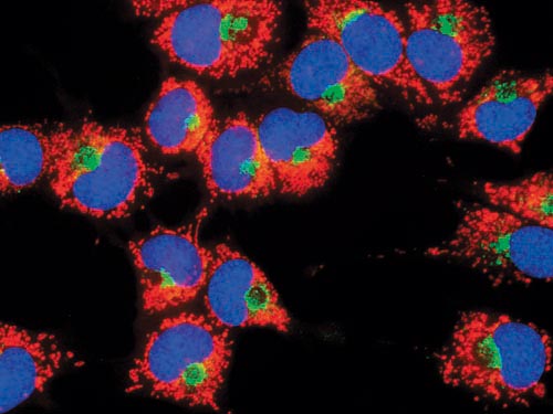
Quantitative and Dynamic Assessment of the Contribution of the ER to  Phagosome Formation - ScienceDirect
Quantitative and Dynamic Assessment of the Contribution of the ER to Phagosome Formation - ScienceDirect

Fluorescence microscope - Wikipedia

Solved Fluorescence microscopy using antibodies tagged with | Chegg.com

Fluorescence microscope - Wikipedia

Fluorescein diacetate 5-maleimide, Fluorescent marker used in microscopy  studies (CAS 150322-01-3) (ab145333)
Fluorescein diacetate 5-maleimide, Fluorescent marker used in microscopy studies (CAS 150322-01-3) (ab145333)

Multi-photon Fluorescence Microscopy - Fluence.technology

Fluorescence Microscopy - Microscopy

Chan Zuckerberg Initiative - A rainbow of colors illuminate the different  cells labeled with fluorescent markers. Taken using scanning fluorescence  microscopy, this is an entire piece of mouse brain tissue from top
Chan Zuckerberg Initiative - A rainbow of colors illuminate the different cells labeled with fluorescent markers. Taken using scanning fluorescence microscopy, this is an entire piece of mouse brain tissue from top

Importance of Colocalization Studies

Newsletter: Fluorophores for Super-Resolution Microscopy - FluoroFinder

Improved Chemical-Genetic Fluorescent Markers for Live Cell Microscopy |  Biochemistry
Improved Chemical-Genetic Fluorescent Markers for Live Cell Microscopy | Biochemistry

Improved Plasmids for Fluorescent Protein Tagging of Microtubules in  Saccharomyces cerevisiae - Markus - 2015 - Traffic - Wiley Online Library
Improved Plasmids for Fluorescent Protein Tagging of Microtubules in Saccharomyces cerevisiae - Markus - 2015 - Traffic - Wiley Online Library

Detection of fluorescent markers by confocal laser scanning microscopy... |  Download Scientific Diagram
Detection of fluorescent markers by confocal laser scanning microscopy... | Download Scientific Diagram
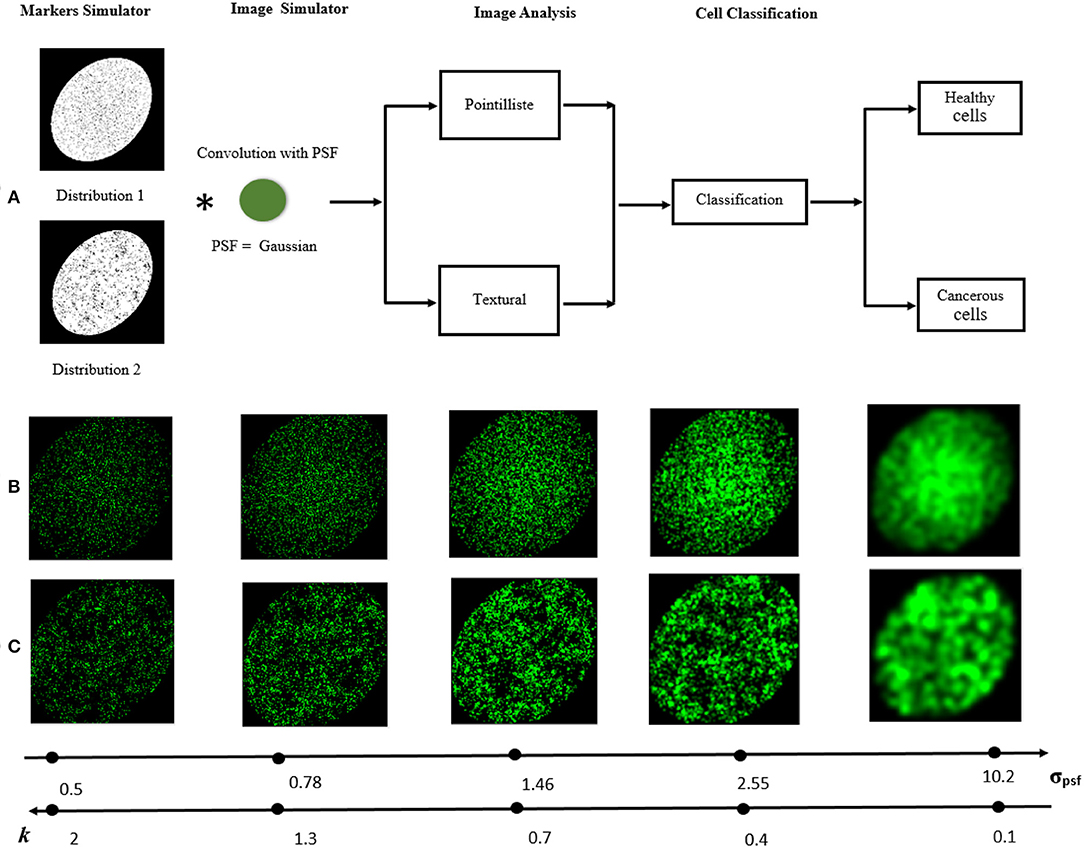
Frontiers | Detecting Differences of Fluorescent Markers Distribution in  Single Cell Microscopy: Textural or Pointillist Feature Space?
Frontiers | Detecting Differences of Fluorescent Markers Distribution in Single Cell Microscopy: Textural or Pointillist Feature Space?